
A toxic protein forms dynamic pores in the membranes of brain cells – and that may be the key to understanding how Parkinson’s disease develops. This is the conclusion of a new study from Aarhus University, where researchers have developed an advanced method to track molecular attacks in real
Molecular movie in slow motion
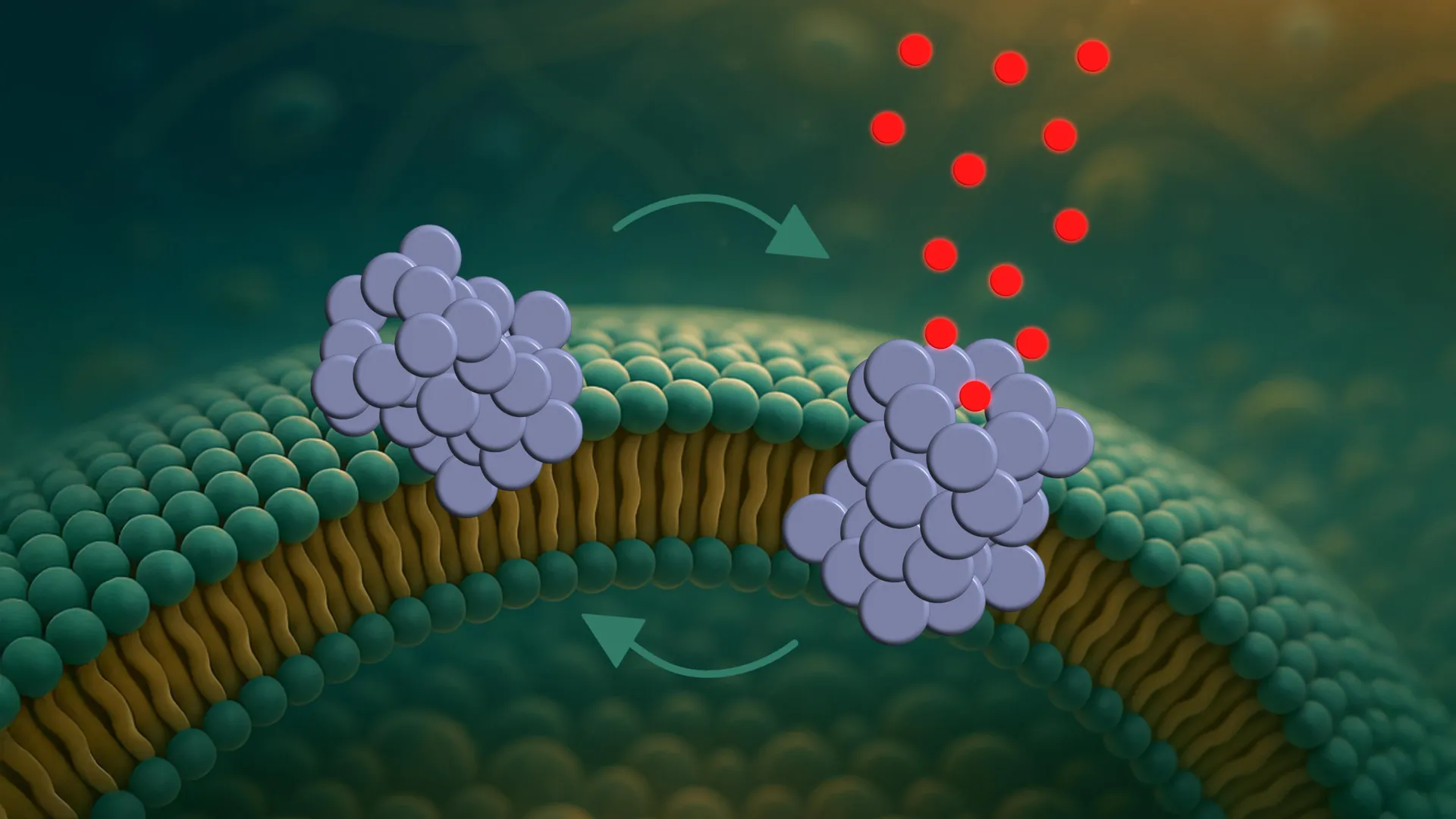
This is the first time such pore dynamics have been observed in real time. It was made possible by a newly developed single-vesicle analysis platform that allows researchers to follow interactions between individual proteins and individual vesicles.
Vesicles are small artificial bubbles that mimic cell membranes and serve as simplified models of real cells.
“It’s like watching a molecular movie in slow motion,” explains Mette Galsgaard Malle. “Not only can we see what happens – we can also test how different molecules affect the process. That makes the platform a valuable tool for drug screening.”
Long road to treatment
Actually, the team has already tested nanobodies – small antibody fragments – developed to specifically bind these oligomers. They show promise as highly selective diagnostic tools.However, as a treatment, there is still some way to go.
“The nanobodies did not block the pore formation,” says Bo Volf Brøchner. “But they may still help detect oligomers at vrey early stages of the disease. That’s crucial, as Parkinson’s is typically diagnosed only after critically important neuronal damage has occurred.”
The study also shows that the pores are not formed randomly.They tend to emerge in specific membrane types – especially those resembling the membranes of mitochondria, the cell’s energy factories. This could indicate that the damage begins there.
Expert Context
Current research suggests that the aggregation of proteins plays a significant role in the development of Parkinson’s disease. The formation of these protein aggregates can disrupt cellular functions, leading to neuronal damage. The ability to observe these processes at a molecular level, as demonstrated by this study, provides valuable insights into the mechanisms underlying the disease. The use of model systems, while offering controlled experimental conditions, necessitates further examination in more complex biological environments to validate the findings and assess their translational potential.
One step at a time
However, the researchers emphasise that the study was conducted in model systems – not in living cells. The next step will be to replicate the findings in biological tissue, where more complex factors come into play.
“We created a clean experimental setup where we can measure one thing at a time.That’s the strength of this platform,” says Mette Galsgaard Malle. “but now we need to take the next step and investigate what happens in more complex biological systems.”
Timeline
- Current: A new study from Aarhus University is released.
- Current: A single-vesicle analysis platform is developed.
- Current: Nanobodies are tested as potential diagnostic tools.
Understanding the dynamics of these protein pores is a crucial step toward developing effective treatments for Parkinson’s disease. The advanced platform developed by the Aarhus University team offers a promising avenue for future research and drug discovery.
Title: Unlocking Parkinson’s Mysteries: Your Nanobody Questions Answered
New research offers fresh insights into Parkinson’s disease by exploring how toxic protein clumps impact cell function.
Q: What are nanobodies and how might they help with Parkinson’s?
A: nanobodies are small antibody fragments designed to specifically latch onto certain protein clumps linked to Parkinson’s. While they haven’t yet shown they can stop pore formation in cells, they hold promise as highly selective tools to detect these clumps very early, which is key since parkinson’s is often diagnosed after meaningful nerve damage has occurred.
Q: did these nanobodies successfully treat the cellular damage?
A: No, the nanobodies did not block the pore formation observed in the study. Though, researchers beleive they could still be valuable for early detection of the protein oligomers.
Q: So, what is the main takeaway about the role of these protein pores?
A: The study has revealed that these damaging pores aren’t formed randomly. They tend to appear in specific types of cell membranes, especially those resembling the membranes of mitochondria, the cell’s energy hubs.
Q: What might the specific location of pore formation suggest?
A: The tendency for these pores to emerge near mitochondria, the powerhouses of the cell, could indicate that the initial damage in Parkinson’s disease begins in these vital organelles.
Q: What type of systems were used in this study?
A: Crucially, this pioneering research was conducted in model systems, not in living cells.
Q: What are the next steps for this research?
A: The researchers plan to replicate these findings in biological tissue.This will allow them to investigate what happens in more complex biological systems, moving beyond the controlled habitat of the lab.
Q: What’s the advantage of the platform used in this study?
A: The platform developed by the Aarhus University team allows for a clean experimental setup, enabling scientists to measure one specific factor at a time. this focused approach is a significant strength for detailed analysis.
Q: Why is understanding these protein pores so important for Parkinson’s?
A: Grasping the dynamics of these protein pores is a critical advancement toward finding effective treatments for parkinson’s disease.
Q: When was this new study released?
A: The new study from Aarhus University is current.
Q: Has a platform for single-vesicle analysis been developed?
A: Yes, a single-vesicle analysis platform was developed as part of this research.
Q: Are nanobodies being tested for Parkinson’s diagnostics?
A: Yes,nanobodies are being tested as potential diagnostic tools in the current research.
This research offers a promising avenue for future studies aiming to decipher Parkinson’s disease mechanisms.